
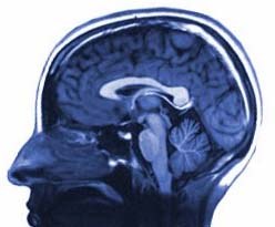
I'm Corree Laule, an MR physicist and Professor at the University of British Columbia. My research bridges advanced MRI techniques with neuropathology to uncover what drives signal changes in human brain and spinal cord tissue.
My work focuses on the microstructural and pathological determinants of MR signal changes. By combining advanced imaging with histological analysis of post-mortem tissue, we develop and validate techniques that provide greater specificity about what's happening in the brain.
In collaboration with neuropathologists Dr. Wayne Moore and Dr. Veronica Hirsch-Reinshagen, we compare MR metrics in post-mortem tissue to histological markers for myelin, axons, and other tissue components — connecting imaging findings to their biological basis.
To interpret abnormalities in disease, we first need to fully characterize healthy tissue. Through MRI studies on healthy controls and NMR experiments on phantoms, we investigate the role of tissue architecture and exchange on T1 and T2 derived measures.
A significant focus of my research. MS attacks myelin — the insulating sheath around neurons. Conventional MRI detects lesions but lacks specificity. We're developing methods that more accurately characterize pathology, helping us understand the mechanisms of damage that lead to disability.
Beyond MS, we develop and apply advanced MR methods across a range of conditions including spinal cord injury, schizophrenia, cervical myelopathy, phenylketonuria, Huntington's disease, brain tumours, dementia, and cerebral malaria.
For the full publication record, visit Corree Laule’s Google Scholar profile or public PubMed bibliography.
I'm very fortunate to work with some amazing people. Our group spans trainees, staff scientists, technologists, and faculty collaborators.
So, how did I end up scanning brains and spinal cords?
The early years….
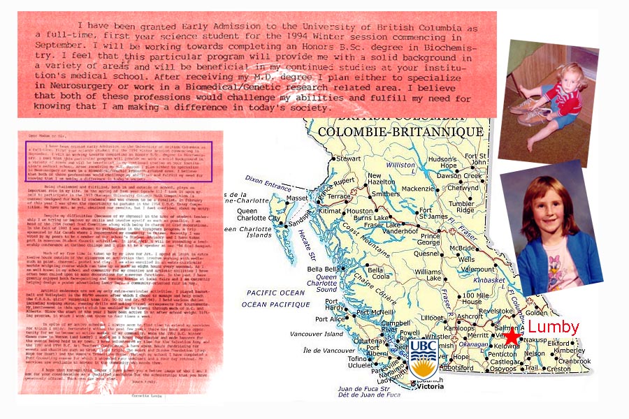
So, off to university I went, excited to begin my career path to becoming a doctor. I was going to help people, I was going to save lives!….And then I took first year biology and discovered that memorizing terms and dissecting a trout were not my cup of tea. I was devastated – if I hated Bio 110, how would I ever become a neurosurgeon?
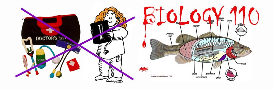
My medical school dreams were put aside as I pondered the question “what will I be when I grow up?”. I’d always liked math and was doing pretty well in physics, so when someone from the fairly new at the time co-op program came into our physics lecture to tell us about how we could take a break from classes and get paid to try out different jobs, I thought it sounded like a pretty sweet deal and signed up.
 Fast foward a few years and I was well on my way to becoming a physicist. I had tried some interesting jobs including ones at NORTEL (not doing so well now!) and TRIUMF, but I didn’t feel passionate about what I was doing. My interest in medicine and biology never really faded, but I was still haunted by the memories of the trout. Then I took a course that changed my life – Introduction to Biophysics. The idea that you could use math and physics to solve biological problems was amazing and something that had never occurred to me. It was the best of both worlds – physics applied to medicine and biology – something practical that could help people! This was also when I first learned about MRI – the idea that you could use giant magnets to take super detailed pictures of the inside of someone’s body without cutting it open was so cool. I was hooked and tried to learn as much as I could about this exciting technology. (Note: The physics department now has a full-fledged Biophysics program, which didn’t exist 15 years ago).
My next co-op jobs involved medical and biophysics, and my honours thesis project was developing an MRI technique to measure myelin in children. I applied to graduate school and received funding from the MS Society of Canada to work on measuring myelin in people with MS. A few years later….
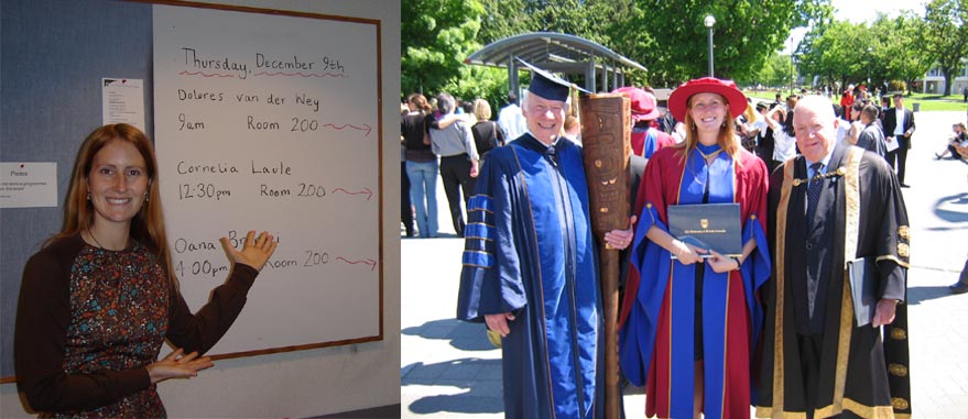
Near the end of my PhD I started working on an MR-histology correlation study that compared a new MR technique we were developing to measure myelin to the ‘gold standard’ histological myelin staining. I found this project very exciting and several years after finishing my PhD in Physics I did a post-doc in the Department of Pathology & Laboratory Medicine. My studies were funded by the Women Against MS Transitional Career Development Award from the endMS Research and Training Network and the Multiple Sclerosis Society of Canada.

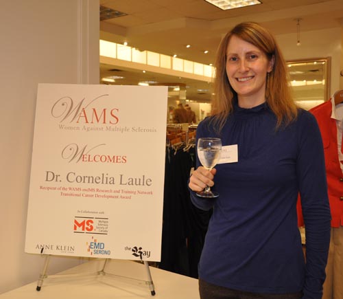
Which brings me to the present. I am currently a Professor in the Departments of Radiology and Pathology & Laboratory Medicine at the University of British Columbia. I’m also an associate member of the Department of Physics & Astronomy. I am Vice Chair, Research in Radiology, Associate Director of Education & Training at the International Collaboration on Repair Discoveries (ICORD) and Director of the UBC MRI Research Centre. I have grant funding from NSERC and the Craig H. Neilsen Foundation. I’m looking forward to continuing my research and updating this page with the next chapter!
And that’s how I ended up scanning brains and spinal cords.